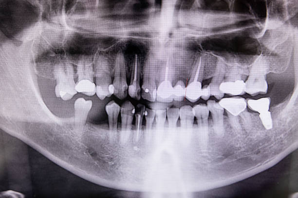
Digital x-ray of teeth profile with root canal, fillings and orthodontic implant at dental clinic
Another significant rhythm is heard in the Dental clinic Memphis, TN where barbecue and the blues meet: the meticulous planning of root canal procedures. In order to save damaged teeth, root canals are necessary, and the periapical X-ray is the unsung hero in this operation. We will examine the critical function periapical X-rays play in Memphis root canal therapy in this blog post, highlighting the significance of this diagnostic tool in maintaining a smile.
A root canal is a type of dental operation used to repair badly damaged or infected teeth. In order to stop the infection from spreading, the dentist first extracts the tooth’s infected pulp, cleans and disinfects the root canals, and then closes the area. Contrary to popular belief, root canal therapy relieves pain brought on by serious tooth decay or infections.
The Importance of Prenatal X-rays
Intraoral X-rays, sometimes referred to as periapical X-rays, are essential to the effectiveness of root canal therapy. These X-rays show the tooth in great detail, including the surrounding bone structure from the crown to the tip of the root. They are essential to the root canal procedure for the following reasons:
- Accurate Diagnosis: Periapical X-rays enable dentists in Memphis to accurately diagnose the extent of infection or decay. These X-rays reveal the roots’ anatomy, the number of canals, and any potential complications, ensuring a comprehensive understanding of the tooth’s condition.
- Treatment Planning: Armed with precise information from periapical X-rays, dentists can create a tailored treatment plan for each patient. This includes determining the appropriate number of root canals to be treated and identifying any additional procedures required for successful treatment.
- Identification of Complications: Some root canals may have intricate structures or unusual anatomy. Periapical X-rays help dentists identify potential complications such as curved canals or extra canals that may be missed during a visual examination alone.
- Post-Treatment Evaluation: After completing the root canal procedure, dentists in Memphis use periapical X-rays to assess the treatment’s success. The X-rays reveal whether the infection has been completely eliminated and the canals are properly sealed, ensuring the best possible outcome for the patient.
Patient Experience and Radiation Safety
Understanding that patients may have concerns about radiation exposure during X-rays, it’s important to note that periapical X-rays involve minimal radiation. Memphis dentists put their patient’s health and safety first. They use the newest equipment and follow tight guidelines to reduce radiation exposure while producing high-quality photos.
Maintaining teeth through quality dental treatment is a common goal in Memphis, where there is a lively pace of life. Periapical X-rays are a dentist’s most useful toolkit, helping them to navigate the complex root canal treatment process with accuracy and gentleness. Memphis residents can be confident that periapical X-rays are used routinely; in fact, they are an essential step in maintaining dental health and making sure that the blues in the music city don’t show up in their smiles.